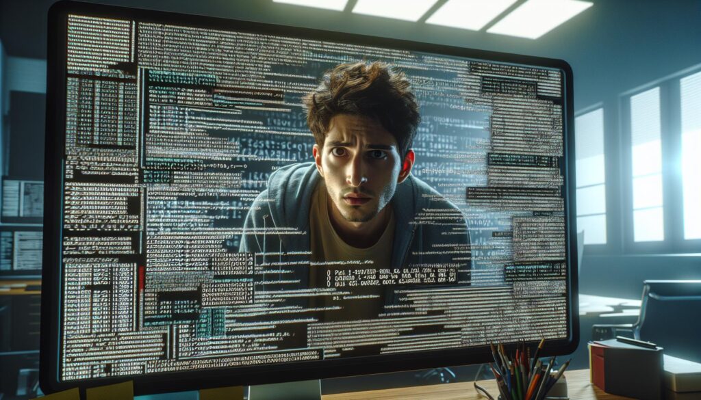
How can a tumour sit there on a scan for two years and no one see it?
It’s much more common than patients understand. A radiologist opens up a scan, sees nothing, signs off the report, and goes to the next patient. Two years later they get another scan which reveals a mass — and the old scan is brought up, and there it is. Subtle. Tiny. Obvious with the benefit of hindsight.
Between “already visible” and “actually noticed” lies where most damage resides.
The good news?
Software is finally starting to close that gap.
What’s covered here:
- Why radiologists miss things in the first place
- What a second-read algorithm actually does
- Where AI catches the most misses
- What this means for medical malpractice claims
Why Radiology Misses Happen So Often
Radiologists are not careless people. They are buried.
A tired hospital reader may go through hundreds of studies per shift. Thousands of images per study. Boredom sets in, focus dims and your eye sees what you THINK you see, not what is actually there.
The statistics prove this as well. One study published in the American Journal of Roentgenology showed that diagnostic errors were responsible for nearly three-quarters of radiology claims. That same study estimated the real-time error rate was approximately 3–5% in routine clinical practice.
Three to five percent doesn’t seem like much. Scale it to millions of scans per year and suddenly it’s very large.
Misses that cause actual harm aren’t clinical problems anymore. They’re legal ones. Mistakes like delayed cancer diagnoses and misinterpreted fractures and missed bleeds top the list of common medical mistakes an attorney handles during a medical malpractice lawsuit. The stakes involved demonstrate how severe those consequences can be: one review of over 6,000 settled claims found the average payout was just under $661,000 for diagnostic errors at doctor’s offices. That’s about twice the amount paid for claims without a clinical error.
Cancer is the most commonly overlooked diagnosis of them all.
What A Second-Read Algorithm Actually Does
Most screening programmes have two radiologists independently interpret the same image. If they disagree, someone else adjudicates. It’s known as double reading. Double reading works – it just requires twice as many staff and twice the money. Most programmes don’t have either.
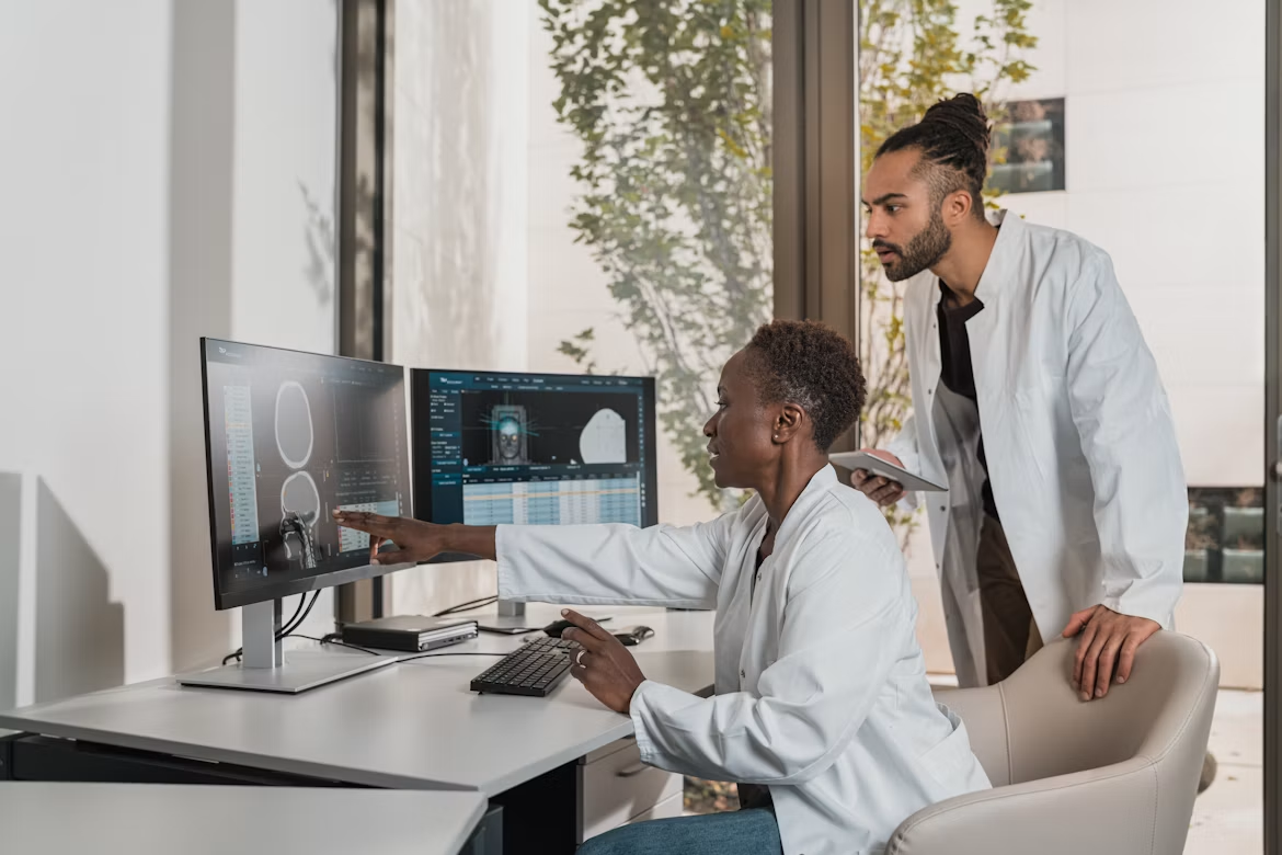
A second-read algorithm steps into one of those chairs.
How it works: The algorithm has learnt from millions of labelled images with a known outcome. As scans are received it will score suspect areas and place an indicator on anything suspicious.
The algorithm does not diagnose anything. It points.
That’s an important distinction. The radiologist always makes the decision, still writes the report and ultimately is responsible. The software just guarantees that a second set of “eyes” has reviewed every pixel without fatigue at 4pm on Friday.
Where AI Catches What Human Eyes Miss
Not every scan benefits equally. Certain areas have shown far stronger results than others.
Breast Screening
This is where the evidence is strongest by a mile.
Large German research involving over 460,000 women participating in an actual screening program revealed AI-assisted reading resulted in a 17.6% increase in cancer detection compared to traditional double reading, without increasing recall rates.
Best of all though, separate data using multireaders found AI-assisted double reading outperformed two humans reading together, achieving 91.8% versus 87.4% sensitivity. The improvement was greatest among lower-volume readers, which shouldn’t surprise anyone.
Then there’s what’s called interval cancer. Those are cancers that develop between screenings. Tumours that take longer to develop tend to be more aggressive. Doctors studying 40,000 mammograms found this added 8.4% sensitivity when AI was used as a second reader. Those were lesions initially passed at the time that later were confirmed as cancerous.
Lung Nodules On Chest Imaging
A small nodule can be obscured on a chest X-ray by a rib, vessel or cardiac silhouette.
Algorithms trained using chest radiographs as well as LDCT will identify these nodules, quantify their size and automatically monitor growth from scan to scan. The ability to measure change over time is the unsung victory. Humans are not good at determining whether or not a nodule grew .1mm since last year. Computers don’t have that problem.
Urgent Findings In The Emergency Department
Approximately 50% of radiology claims originate from ED visits. This is understandable considering the hectic nature of the ED.
Triage algorithms look for life-or-death findings such as intracranial hemorrhage, massive stroke, pneumothorax, and move those studies to the front of the queue. The radiologist still reads the study. They just read it in eight minutes rather than two hours.
What This Means For Medical Malpractice
Here’s where things get interesting.
Each AI flag leaves a record behind. If code flags an area and the report written contains no mention of it, that flag still remains ~ stored with a time stamp ~ for whomever cares to search for it later.
That cuts in a few directions:
- A cleaner paper trail. Reviewers know exactly what the software reviewed and when it reviewed it.
- A moving goalpost. “Everybody misses stuff” stops being a good defence as this becomes commonplace.
- Fresh doubts about dependability. Who is responsible when the algorithm doesn’t speak and the reader depends on that silence?
None of this is fully resolved, either in courts or with regulators. However, you can see where this is going. Documentation is improving, which changes how you build a medical malpractice case.
Where The Technology Still Falls Short
Nobody should oversell this.
More flags equals more false positives. False positives equal more callbacks, more biopsies and more anxious patients waiting for results that are benign.
And of course automation bias. If you train your reader to trust your green light they will want to skim rather than inspect. That is a very real danger, and completely counter to what the tool is intended to do.
Also performance is all over the place between vendors. Because a tool was validated on one population does not mean it will work on another population. Cleared for market != proven in your hospital.
The Bottom Line
Second-read algorithms are not replacing radiologists. Never will be. They pick up what the exhausted human eye glazes over – that subtle nodule, that 3mm progression, that cluster of calcifications you didn’t think much of at the time.
Quick recap:
- Radiology misses are common, and cancer tops the list
- Second-read algorithms flag suspicious regions, they don’t diagnose
- Breast screening shows the strongest results so far
- Every flag creates a permanent record
- The technology helps, but it isn’t a substitute for careful reading
Think about how that applies to patients. If you get a diagnosis too late when damage has already occurred, you want to know what did the imaging actually reveal — and was anyone, person or computer, watching?