When vertical bone height limits your options and you want to avoid grafting, short dental implants in Greenville, SC give you a practical, less invasive path to restore missing teeth. Short implants can often replace longer fixtures without bone augmentation, offering high survival rates and fewer complications in many posterior sites.
You will learn when short implants suit your case, how implant design and biomechanics affect success, and how outcomes compare with traditional graft-and-implant approaches. Expect clear guidance on clinical indications, long-term maintenance, and what to discuss with your clinician before choosing a graftless solution.
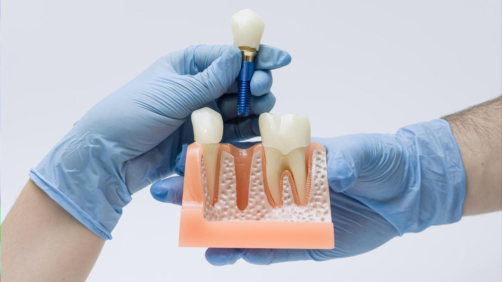
Clinical Indications for Short Implants
Short implants let you avoid vertical grafting, reduce treatment time and cost, and still achieve predictable support in sites with limited bone height or where augmentation is contraindicated.
Assessment of Residual Bone Height
Measure vertical bone height from crest to vital anatomy (maxillary sinus floor or inferior alveolar nerve) using CBCT. You need at least the planned implant length plus a safety margin—commonly 1.5–2.0 mm to the nerve and 1.0–2.0 mm to the sinus floor.
Evaluate crestal bone width and cortical quality as well; a narrow ridge may require simultaneous ridge expansion or a wider-diameter short implant.
Record existing bone density (D1–D4); denser bone improves primary stability, which matters more with reduced implant length.
Plan for prosthetic needs: occlusal scheme, crown-to-implant ratio, and anticipated lateral loads. If crown height exceeds 15 mm or parafunction exists, reconsider loading strategy or splinting.
Patient Selection Criteria
Choose short implants when the patient refuses or cannot undergo grafting due to medical comorbidity, smoking, or financial/time constraints.
Ideal candidates have localized vertical deficiency but adequate horizontal bone and good oral hygiene. Manage expectations about potential need for occlusal adjustments and maintenance.
Contraindications include uncontrolled systemic disease, active infection at the site, very thin cortical plates, or extreme parafunction without protective measures.
Prefer short implants for older patients or those seeking less invasive care, and for posterior sites where splinting to adjacent units is feasible to distribute forces.
Case Examples in Posterior Regions
Posterior maxilla with 6–8 mm of residual height: place 6–8 mm short implants to avoid sinus lift when CBCT shows adequate width and at least 1–2 mm clearance from sinus. Use platform-switching and roughened surfaces to enhance bone response.
Posterior mandible with 7–9 mm height above the nerve: select short implants of 6–8 mm and aim for bicortical engagement if possible. Maintain a 1.5–2.0 mm safety distance from the inferior alveolar canal.
When multiple adjacent teeth are missing, splint short implants across the edentulous span to reduce cantilever forces. Adjust occlusion to minimize lateral loads and consider nightguard therapy for bruxers.
Biomechanical Considerations and Implant Design
Short implants rely on optimized shape, diameter, and surface to transfer loads predictably into limited-height bone while avoiding grafting and additional surgery.
Load Distribution and Prosthetic Planning
You must plan occlusion and prosthetic design to reduce lateral forces on short implants. Place contacts in centric relation and minimize nonaxial loading through careful occlusal scheme adjustments.
Angulation and implant position affect bending moments. Where possible, align the implant long axis with the occlusal load vector and consider splinting adjacent implants to distribute forces across multiple fixtures.
Use prosthetic components that reduce cantilevers. Short crowns and controlled crown-to-implant ratios lower moment arms. Consider using occlusal materials with some shock absorption to dampen peak forces.
Implant Length Versus Diameter
When height is limited, increasing diameter often improves primary stability and bone contact area more effectively than increasing length. A wider implant increases surface area at the crestal bone, improving load transfer in Type II–IV bone.
Balance diameter with local anatomy. Sinus proximity, interradicular width, and cortical thickness constrain implant diameter choices. Choose the largest diameter that preserves at least 1.5–2 mm of bone between implant and adjacent structures.
Thread design and core geometry influence mechanical engagement. Deeper threads and progressive tapering can boost insertion torque and initial stability without increasing length.
Surface Modification Technologies
Surface roughening and bioactive coatings accelerate osseointegration and increase bone-to-implant contact, which matters more for short implants because contact area is inherently limited. Use implants with proven micro-rough or nano-structured surfaces.
Consider surfaces that promote early stability and bone apposition, such as controlled grit-blasting plus acid-etch or calcium phosphate-based coatings. These surfaces shorten healing time and help support earlier loading protocols.
Pay attention to platform switching and connection type. Conical connections and platform switching can reduce crestal bone stress and micro-movement, preserving marginal bone around short implants.
Comparative Outcomes and Complication Rates
Short implants often match standard-length implants for survival while showing lower rates of some complications. You can expect similar prosthesis success with less need for bone grafting and fewer biological complications in many published trials.
Success and Survival Rates
Randomized controlled trials and meta-analyses report survival rates for short implants (commonly ≤8.5 mm, with many studies on ≤6–7 mm) that are comparable to standard implants placed with grafting.
You should expect implant survival commonly above 90–95% at 3–5 years in posterior maxilla/mandible cases when implants are well planned and placed in adequate residual bone.
Key practical points:
- Short implants avoid donor-site morbidity and reduce surgical time compared with sinus lifts or vertical augmentation.
- Survival parity is most consistent when you use modern implant designs (wider diameter, improved surface) and follow prosthetic guidelines that minimize off-axis loading.
- In cases with extreme occlusal forces, parafunction, or very poor bone quality, survival differences may emerge in favor of augmented longer implants; evaluate case-by-case.
Marginal Bone Loss Analysis
Multiple reviews show that short implants frequently exhibit equal or less marginal bone loss (MBL) than longer implants with grafting.
You will often see reduced MBL near short implants, especially when prosthetic loading protocols and occlusal schemes are controlled.
Factors that influence MBL:
- Implant diameter and platform design — wider platforms and platform-switching reduce crestal bone remodeling.
- Prosthetic factors — crown height, occlusal contacts, and cantilevers increase MBL risk.
- Surgical technique — flapless placement and atraumatic handling can limit early bone loss. Monitor peri-implant bone levels radiographically at baseline, 6–12 months, then annually to detect progressive changes early.
Risk Factors for Implant Failure
You must assess patient, site, and prosthetic variables that raise failure risk with short implants.
Common risk enhancers include poor bone quality (type IV), uncontrolled bruxism, heavy occlusal forces, active smoking, and inadequate implant diameter relative to load.
Practical mitigation strategies:
- Favor larger-diameter short implants where vertical space limits length.
- Use splinting for multiple adjacent short implants to distribute occlusal load.
- Optimize prosthetic design: avoid long cantilevers, reduce crown height when possible, and ensure passive fit.
- Manage systemic and local risk factors before surgery: smoking cessation, glycemic control, and treatment of active periodontal disease.
Long-Term Maintenance and Patient Education
Maintain regular surveillance, control plaque effectively, and attend scheduled maintenance visits to protect bone levels and implant function. You should understand signs of tissue inflammation, follow precise home-care techniques, and commit to professional cleaning intervals tailored to your risk factors.
Peri-Implantitis Prevention
Peri-implantitis risk rises with plaque accumulation, smoking, and uncontrolled diabetes. You should get a baseline radiograph after prosthesis delivery and periodic radiographs (typically annually) to detect early crestal bone changes.
Schedule professional maintenance every 3–6 months based on risk assessment. During visits, clinicians will remove biofilm with titanium- or plastic-tipped instruments, assess probing depths, and record bleeding-on-probing and suppuration. If you smoke, quit or reduce tobacco use; even light smoking increases failure risk.
Control systemic contributors: keep blood glucose within target ranges and review medications that affect healing. If inflammation appears, escalate care promptly—local debridement, antiseptic rinses (0.12%–0.2% chlorhexidine short term), and adjunctive systemic antibiotics only when indicated by your clinician.
Oral Hygiene Recommendations
Clean around short implants twice daily with low-abrasive fluoride toothpaste to protect prosthetic surfaces. Use an interdental brush sized to the embrasure and a soft nylon-coated brush for abutment areas; metal brushes can scratch titanium and trap bacteria.
Daily flossing with implant-specific floss or a floss threader under fixed restorations removes submarginal plaque you cannot reach with a brush. Consider a water flosser set to moderate pressure to dislodge debris without damaging peri-implant tissues.
Use antimicrobial mouthrinses short-term after surgical procedures or during flare-ups as advised. Replace interdental brushes and floss every 1–2 weeks and bring your home-care devices to maintenance visits for technique review and reinforcement.